
NIT Calicut’s smart microscope puts AI to work at the doctor’s eye
KOZHIKODE: What if a microscope could do more than magnify what lies beneath its lens?
KOZHIKODE: What if a microscope could do more than magnify what lies beneath its lens?
Article outline
- What happened
- Why it matters
- The details
- Official response
- The bottom line
Key points
- One of the most significant features is that the system does not depend on an internet connection or cloud-based computing.
- This edge-AI approach could prove particularly valuable in healthcare settings where sophisticated computing infrastructure is not readily available.
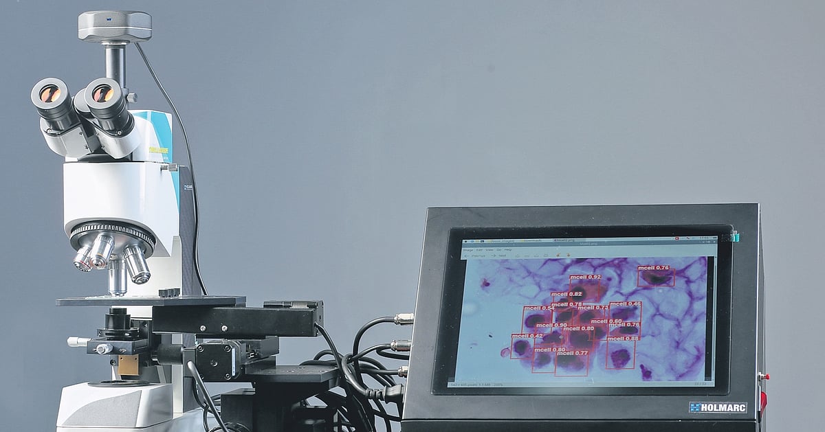
- At the heart of the system is a compact combination of a digital camera, a lightweight AI processor and an LCD.
- The project brings together expertise from engineering, artificial intelligence and medicine.
KOZHIKODE: What if a microscope could do more than magnify what lies beneath its lens? As a medical professional scans a slide, artificial intelligence could quietly pick out suspicious cells, flag areas that deserve attention, and present the findings instantly on a screen?, what if.
That possibility is now taking shape at the National Institute of Technology Calicut, where researchers, working alongside medical experts, have developed an AI-enabled smart microscopy system designed to turn conventional microscopes into intelligent medical imaging platforms.
For context, the technology is aimed at tackling one of the less visible challenges in pathology – the time, concentration, and visual effort required to examine microscopic samples manually. Instead of requiring doctors and pathologists to spend long periods peering through microscope eyepieces, the system captures images or video from the microscope and analyses them in real time, highlighting cells and other features of interest.
At the heart of the system is a compact combination of a digital camera, a lightweight AI processor and an LCD. While an AI model processes the images locally, the camera captures the microscopic field. The results are then displayed on the screen with highlighted regions of interest and visual annotations, allowing medical professionals to examine the material more conveniently.
One of the most significant features is that the system does not depend on an internet connection or cloud-based computing. The AI processing takes place directly on the device, enabling rapid, low-latency analysis while keeping the equipment compact and relatively cost-effective.
This edge-AI approach could prove particularly valuable in healthcare settings where sophisticated computing infrastructure is not readily available. While the technology could be deployed in major hospitals and high-volume laboratories, its compact design additionally raises the possibility of apply in primary healthcare centres and facilities in resource-constrained settings.
Dr Jayaraj P B, associate professor, NIT Calicut. This person led the team, talking regarding the invention, stated, "Our team first focused on urine cytology screening, particularly the identification of cells and microscopic features relevant to urinary bladder cancer screening".
But the researchers see possibilities far beyond urine samples. With suitable training, the underlying imaging and AI architecture could be adapted for cervical cytology, blood smears, histopathology slides and a range of other cytology and pathology applications, he continued.
Dr Jayaraj further remarked, "By bringing AI closer to the microscope itself, our team is trying to make medical image analysis faster, less laborious and more consistent – while keeping the final clinical judgement firmly in the hands of medical professionals."
In practice, the project brings together expertise from engineering, artificial intelligence and medicine. It was backed under the Scheme for Promotion of Academic and Research Collaboration (SPARC) of the Ministry of Education, Administration of India. The technology is now being positioned for a journey beyond the laboratory. Its commercialisation is planned through HealForge AI Private Limited, a firm incubated at the Technology Business Incubator (TBI), NIT Calicut. For context, the objective is to translate the research prototype into a deployable AI-assisted microscopy solution for clinical and laboratory employ.
Meanwhile, the inventors behind the system are Arathy Menon N P, research scholar, NIT Calicut; Dr Jayaraj P B, associate professor, NIT Calicut; Dr Pournami P N, associate professor, NIT Calicut; Dr Simi S, assistant professor, department of pathology, Administration Medical College, Kozhikode; and Dr Swathy Shanker, assistant professor, department of pathology, KMCT Medical College, Kozhikode.
For now, NIT Calicut's smart microscope puts AI to work at the doctor's eye remains the part of the story worth watching, and further updates are likely as more details are confirmed.